Since the Nobel Prize (Binnig and Rohrer, 1986), scanning tunneling microscopy has developed into the decisive experimental tool for the nanosciences.
The basis for the functioning of a scanning tunneling microscope is the quantum mechanical tunneling effect. If two electrically conductive electrodes are brought together so closely that they are separated only by a thin insulating layer, for example by a vacuum, an overlap of the wave functions occurs and electrons can tunnel through the barrier from one electrode to the other. If a voltage of approximately U ~ 1 V is applied between the electrodes, a macroscopic current of the order of nA is measurable. In the scanning tunneling microscope (STM), one of the two electrodes is realized by a fine tip, which functions as a probe. The radius of the tip is typically 20-500 nm and is approximated to about 0.1-0.5 nm to the surface of a sample that is the second electrode of the tunneling contact. Due to the strong dependence of the tunneling current on the distance between the tip and the sample, the surface of a sample can be examined in atomic resolution by scanning the tunneling current as a function of the location.
The following relation applies:
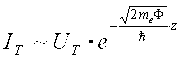
Due to the exponential relation between tunnel current and distance, a very high z-resolution is achieved. In our case it is about 1 pm (!). Therefore, almost the entire tunneling current also flows over a next top atom to the surface.
In essence, a scanning tunneling microscope consists of a scanning unit and a control electronics unit. In both scanning tunneling microscopes used, the scanning unit is implemented as a so-called tube scanner, on which the STM tip is fixed. The tube consists of a piezo crystal in the form of a tube, which has an internal electrode and an external electrode divided into four quadrants. By applying an electrical voltage between the inner and all four outer electrodes, the tube is stretched in length and the tip is positioned in the z direction. However, the four outer electrodes can also have different voltages to the internal electrode. Thus one side of the tube can be stretched and the other side compressed, whereby the tube can be bent and the tip can be accurately positioned in the x- and y-directions to the fraction of an atomic diameter.
The measurement of the sample topology can be done in two different ways:
1.) With the constant height mode, the z-position of the tip is kept constant and the sample is scanned line-by-line in the x- and y-direction. Differences in height on the sample surface vary the tunneling current and is measured as a function of the location. For the representation of the sample topology, the tunneling current is translated into brightness values and displayed as a brightness contrast in an image. The advantage of this method lies in the achievable high scanning speed, so that with an image refresh rate of more than 25 images per second can be measured. However, this mode is only suitable for flat surfaces, since there is a risk of contact with the sample surface.
2.) Another method is the constant current mode. In this case, the tunneling current is kept constant by controlling the distance between the sample and the tip during the rastering over the sample surface, while the z movement of the scanner is measured as a function of the location. These altitude information can be translated as above into brightness values and the topography can thus be displayed in a picture. This mode is the most common mode of operation of scanning tunneling microscopes and provides a higher vertical resolution than the constant height mode. However, the achievable scanning speed in this mode is much lower than in the constant-height mode since the z position of the scanner is constantly controlled and regulated by a control circuit. Recording an STM image usually takes between a few minutes and several hours.
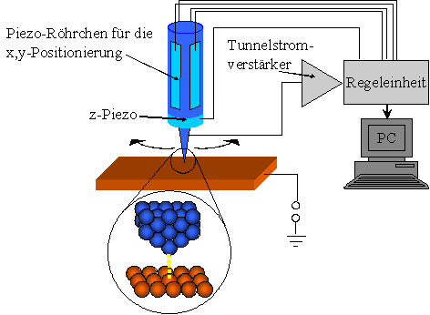
Schematic structure of a scanning tunneling microscope
Our work group has two scanning tunneling microscopes: an Omicron micro-STM (room temperature), as well as a helium-cooled low-temperature STM (Omicron LT-STM).